


BSD4343 Wired


BSD4343 Wired
Description
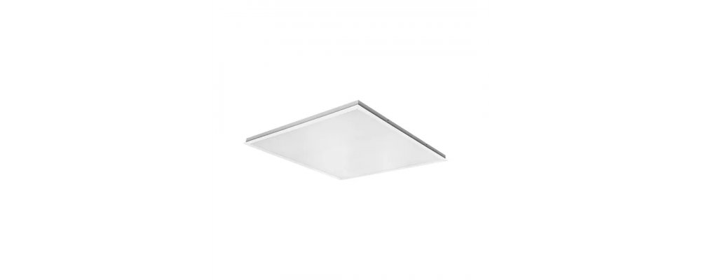
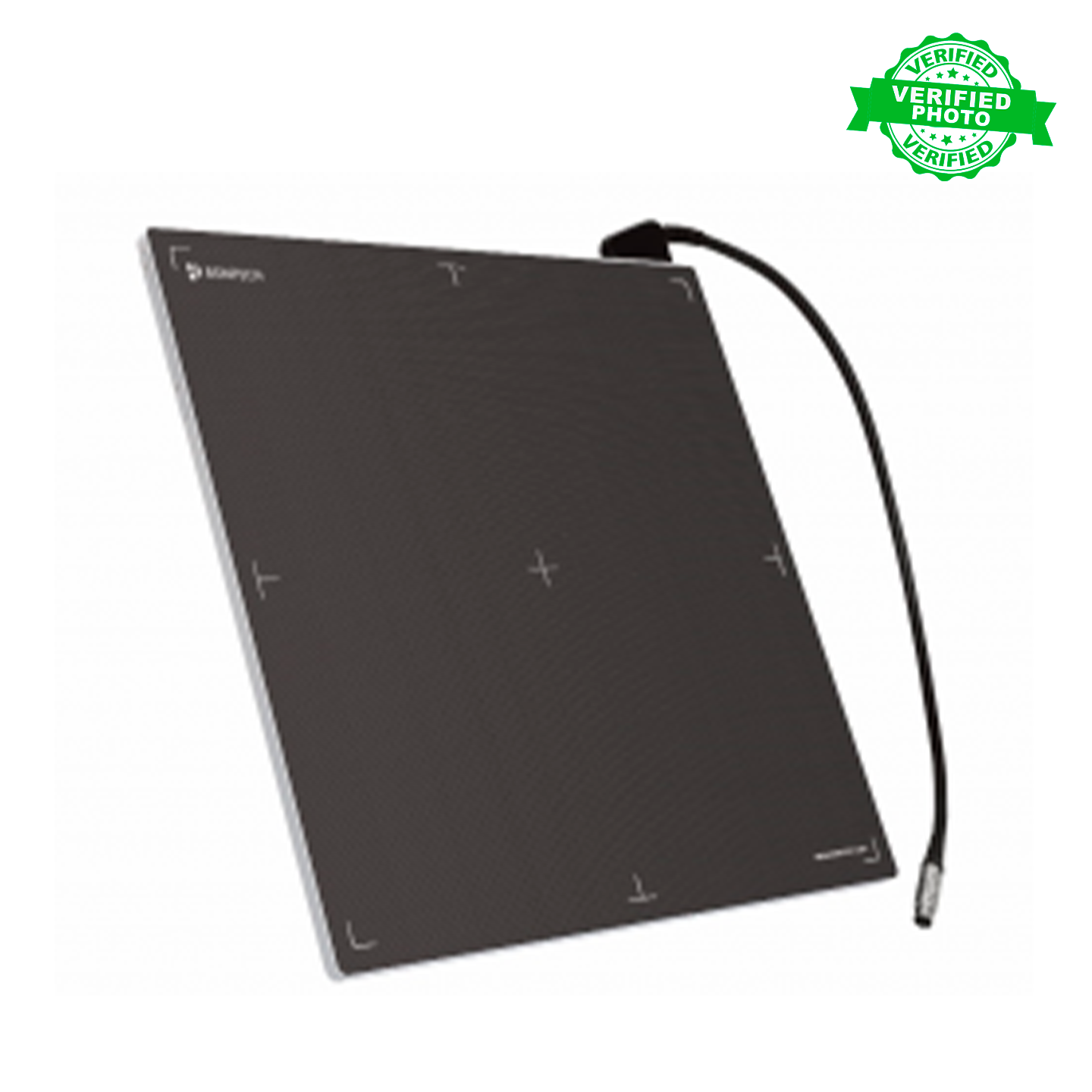
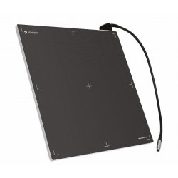
Portable Flat Panel Detector for Digital Radiography Stable and Reliable AED (Automatic Exposure Detection) Bontech Medical's own AED function with reliable performance Support line trigger mode High image quality Wide active area 430.08 x 430.08 High spatial resolution 140m
Characteristics Purpose: General radiography Fill factor: 60% Image matrix size: 3072 x . Power supply: input: 100-240 V, 47-3072 63 Hz, output: 12 V, 2.5 A. Pixel Pitch: 140 µm Interface: Gigabit Ethernet (1000BASE- Effective Area T) via PoE Imaging: 430.08mm x 430.08mm Dimensions: 460mm x 460mm x 15mm. Grayscale image: 16 Weight: Approximately 5 kg without handle bits, 65,536 grayscale: . Scintillator: Csl, GOS Image Acquisition and Time Components The BSD4343(BT-DA24-IA/BT-DB24-IA) has the following components: Flat Panel X-ray Detector (Csl: BT-DA24-IA, Gdos: BT-DB24-IA) Control Box (BT-CBO2) Switching Adapter (W-SAO2) AC Power Cord (BT-SCO1) Mains Cable (BT-ECO2) X-Ray Switching Signal Cable (Sync Cable) (BT-TCOS) Extended Cable (BT-MC05) General The BSD4343 (BT-DA24-IA / BT-DB24-IA) is a cassette-type wired detector for versatile use with excellent image quality. The BSD4343 (BT-DA24-IA / BT-DB24-IA) provides large x-ray images that maximize the anatomical field of view with a minimum of x-rays. Images transferred from this system to RawlmageViewer (BT-IVO1) are processed and transferred to printers for film output or image servers for storage instead of conventional film system radiographic images. The BSD4343 Medical Imaging System (BT-DA24-IA / BT-DB24-TA) is designed to create radiographic images of the human anatomy. The Digital X-Ray Detector, which uses a large-area amorphous silicon matrix sensor, allows X-ray images to be taken without the use of a conventional film/screen system. The BSD4343 (BT-DA24-IA / BT-DB24-IA) is a flat panel X-ray detector used for radiological applications, part of a digital imaging system. The BSD4343 detector (BT-DA24-IA / BT-DB24-IA) is an X-ray imaging device based on a flat panel. The digital X-ray detector uses a large area amorphous silicon array with a CSI or Gadox scintillator. The 350x427.28mm panel will display high quality images for 3 seconds over a wide range of rate settings. This device must be integrated with a workstation [PC] and an X-ray generator. It can be used to digitize x-ray images and transfer for radiographic diagnosis. In addition, it is designed to replace the X-ray film or screen system in all general diagnostic procedures. processing to create a printed image. A device called a detector panel is used to capture images electronically.
No reviews found